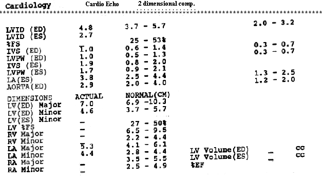
Clinical indication: recurrent pulmonary edema. Known diastolic dysfunction. This is an excellent study. The aortic valve leaflets are thickened with fairly well preserved leaflet mobility. Forward flow velocity is increased with a calculated peak gradient of 33 mmHg and a mean gradient of 21 mmHg. The increased forward flow velocity is probably due to the mild to moderate aortic regurgitation. The left atrium is normal size. The mitral leaflets are slightly thickened and there is calcification seen in the region of the mitral annulus. Forward flow velocity is characterized by almost equal E and A waves and normal pulmonary venous flow pattern is seen. The deceleration time is 160 msec. peak indication marked improvement in the previously prolonged deceleration time indicative of diastolic dysfunction. The left ventricle shows concentric hypertrophy with well preserved systolic function. Ejection fraction is well over 60-70%. Right sided chambers are normal size. The tricuspid valve morphology is normal. Tricuspid forward flow suggests diastolic compliance abnormality of the right ventricle with a pattern suggesting diminished passive filling. A trace of tricuspid regurgitation is present with a right ventricular systolic pressure estimated at 49mmHg plus the right atrial pressure. The pulmonic valve appears normal. IMPRESSION: SINCE THE PRIOR STUDY OF 12-23-93, THERE HAS BEEN FURTHER IMPROVEMENT IN THE DECELERATION TIME, AND THE FINDINGS OF POSSIBLE RIGHT VENTRICULAR DIASTOLIC DYSFUNCTION HAVE APPEARED. THE AORTIC GRADIENTS, DEGREE OF AORTIC REGURGITATION, AND THE RIGHT VENTRICULAR SYSTOLIC PRESSURE ARE SIMILAR ON THE PRESENT STUDY TO THE PRIOR STUDY. | 